
A Woman with Double Chamber Right Ventricle
Article Sidebar
Main Article Content
Abstract
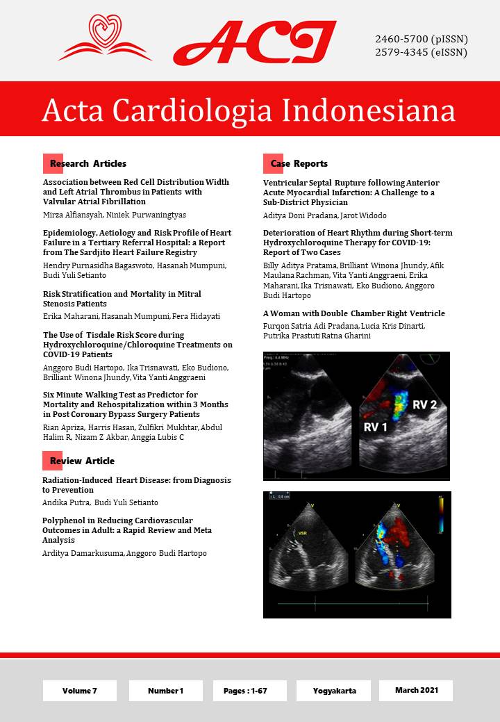
Introduction: An additional membrane or muscle band inside right ventricle divides it into two chambers, the proximal and distal one. It is a rare congenital malformation which makes up approximately 0.5-1 per cent of all congenital heart defects. The double chambers right ventricle most often present in children but rarely in adults. Transoesophageal Echocardiography is the most effective tool in diagnosing a DCRV.
Case presentation: A 36 years old woman, diagnosed with large ventricular septal defect by transthoracic echocardiography, underwent a TOE to complement the preparation of surgical closure. The TOE finding instead revealed a septum or membrane that divide the right ventricle into two separate chambers, later confirmed by a cardiac computer tomographic scan and cardiac cathetherization. The patient underwent surgery with VSD closure and resection of the septum without complications. Echocardiography paramater became normal albeit still had some septal tissue left in order to limit sudden surge of blood flow into pulmonary artery. Follow up indicate patient has no symptom associated with the surgery
Conclusion: The patient was diagnosed with double chamber right ventricle during preparation for surgery by TOE examination. We present this case in order to emphasize the rarity of this congenital heart disease